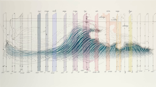
Sleep appears in the brain as Slow waves rising It crossed the surface at a rate of about one every tenth of a second – or so we thought.
A new study in mice suggests that there are patterns of brain activity associated with sleep that we have overlooked — which reflect the state of individual brain cells rather than the collective activity of millions or billions of neurons.
Furthermore, when measuring these highly localized, millimeter-scale brain signals using single-wire electrodes, the researchers found that parts of the mammalian brain may fall asleep for short periods of time while other regions remain broadly awake.
“It was surprising to us as scientists to find that different parts of our brains actually take short naps while the rest of the brain is awake.” He says “This is a very important finding,” said David Haussler, a bioinformatician at the University of California, Santa Cruz, and lead author of the study.
For a century or so, patterns of electrical activity throughout the brain have been used to quantitatively determine the difference between sleep and wakefulness. These brain waves are often detected using electroencephalogram (EEG) by electrodes placed on the scalp.

But Haussler and his team wondered how we measure sleep and distinguish it from wakefulness — when there is clearly some overlap in the brains of animals that stay awake during sleep, a skill known as Unihemispheric slow-wave sleep.
In the 1960s, researchers first suspected and then discovered how Dolphins and other cetaceans They can rest half of their brains while staying active, sometimes keeping one eye open to watch for predators and maintain contact with others in their group.
Seals and birds also exhibit different forms of this rest that alternate between sleep and wakefulness—a clever trade-off between sleep and survival.
Humans can also temporarily exhibit asymmetric sleep patterns that are reminiscent of, but not identical to, those seen in animals.
In 2016, researchers at Brown University in the United States found that the first night people sleep in an unfamiliar place, the left side of the brain is active. More alert to deviant sounds From the right. Once we get used to the sleeping environment, this difference disappears.
“The human brain has been shown to have a less dramatic form of monosomic sleep than that found in birds and some mammals,” says neuroscientist Christof Koch. books in Scientific American Magazine When those results were published.
If the mouse brain is the standard, then the lack of clarity of wake and sleep states in humans may be a neural trait we share with other animals after all.
Haussler and his team collected data over weeks from nine mice that had thin electrodes implanted in 10 different areas of their brains, then fed that data into an artificial neural network that learned to distinguish between sleep and wake states.
Recordings were sampled from 100 micrometers (one-tenth of a millimeter) of brain tissue, and the algorithm was able to reliably identify sleep-wake cycles based on brief “flashes” in brain cell activity lasting only 10 to 100 milliseconds.
These “local” signals indicate that part of the animals’ brains were asleep while other areas remained active and awake. Coincidentally, the researchers noted, this occurred at a time when a mouse might have stopped moving for a fraction of a second, as if it had “lost consciousness.”
“We were able to look at individual time points when these neurons fired, and it was very clear that [the neurons] “They were in transition to a different state.” It is clear “This is a very interesting study,” said Aiden Schneider, a computational biologist at Washington University in St. Louis, who co-led the study with David Parks, a graduate student in computer science at the University of California, Santa Cruz.
“In some cases, these flashes may be limited to just one area of the brain, and may be smaller than that.”
The team believes their new method of measuring sleep and wake states could reveal new secrets about how we sleep, if other research groups can observe these “flashes.”
“they [the flickers] “Break the rules you would expect based on a hundred years of literature,” He says Neuroscientist Keith Hengen of Washington University in St. Louis.
The study was published in natural neuroscience.

“Typical beer advocate. Future teen idol. Unapologetic tv practitioner. Music trailblazer.”


More Stories
Boeing May Not Be Able to Operate Starliner Before Space Station Is Destroyed
How did black holes get so big and so fast? The answer lies in the darkness
UNC student to become youngest woman to cross space on Blue Origin